ONE-STOP DREAM SOLUTION
Anterior Segment
ONE-STOP DREAM SOLUTION
Anterior Segment
The DREAM OCT™ could acquire the corneal curvatures for both anterior and posterior surfaces, the topography of corneal epithelium, the axial length, AS-OCT images and associated measurements on only one device, providing accurate and reliable data for refractive surgeries, contact lens treatment, and ICL implantation.
Screening before Refractive Surgeries
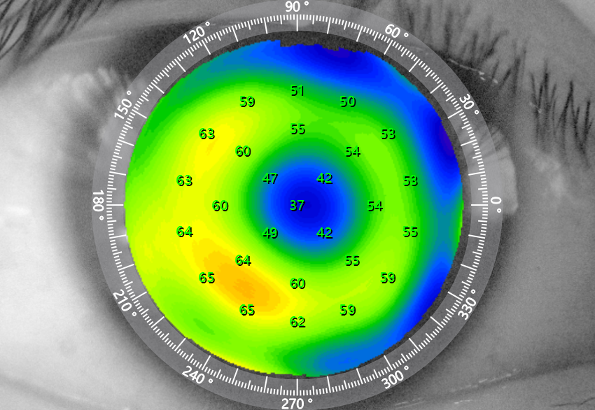
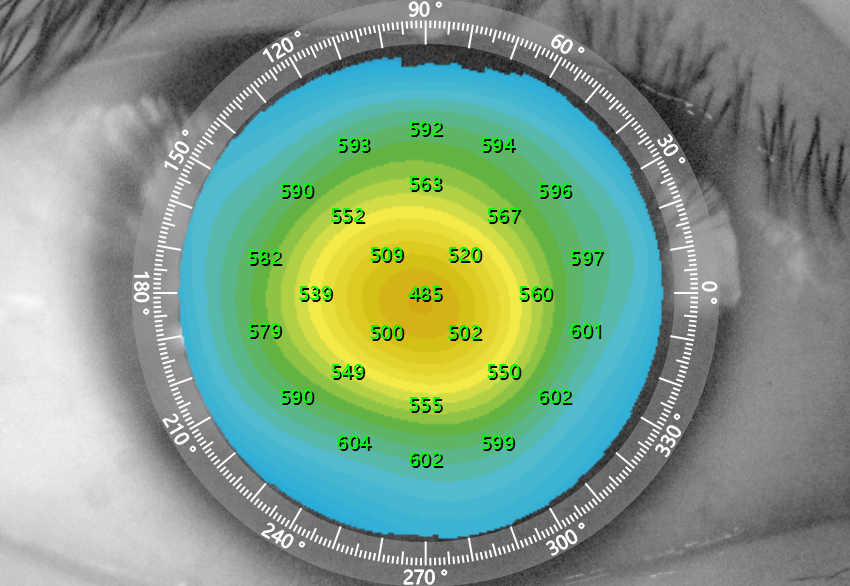
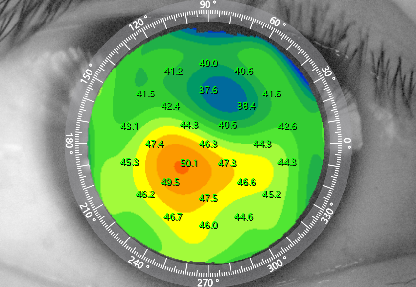
The DREAM OCT™ could provide not only the corneal thickness topography, the corneal curvature diagram, the corneal height diagram for both anterior and posterior surfaces, but also the corneal epithelial thickness topography. So that we could do comprehensive screening for corneal condition, which is very important for the assessment before refractive surgeries.
Corneal epithelial thickness topography
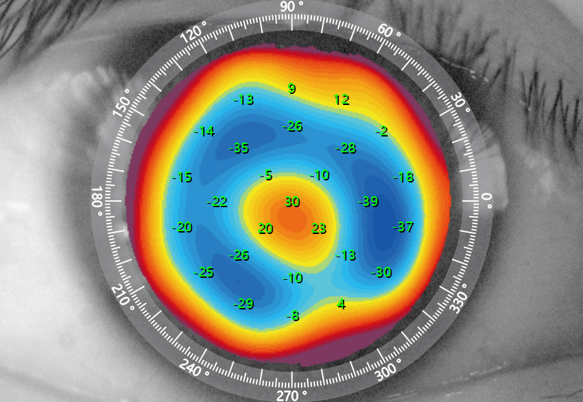
Corneal height diagram of posterior surface
Corneal thickness topography
Corneal curvature diagram
Higher Success Rate of Axial Length Measurements for Patients with Ocular Media Opacity
The DREAM OCT™ could penetrate severe opacity of lens much more easily, and measure the axial length precisely based on the visualized OCT imaging from anterior segment to posterior segment along the visual axis, providing reliable data for IOL calculation before surgery.
AI Guided Automatic Measurements for AS-OCT
Using AI technology, the software could automatically identify many structures such as the scleral spur, and measure the anterior chamber depth, ICL vault, corneal flap thickness, etc., in order to assess the effect of refractive surgeries objectively.
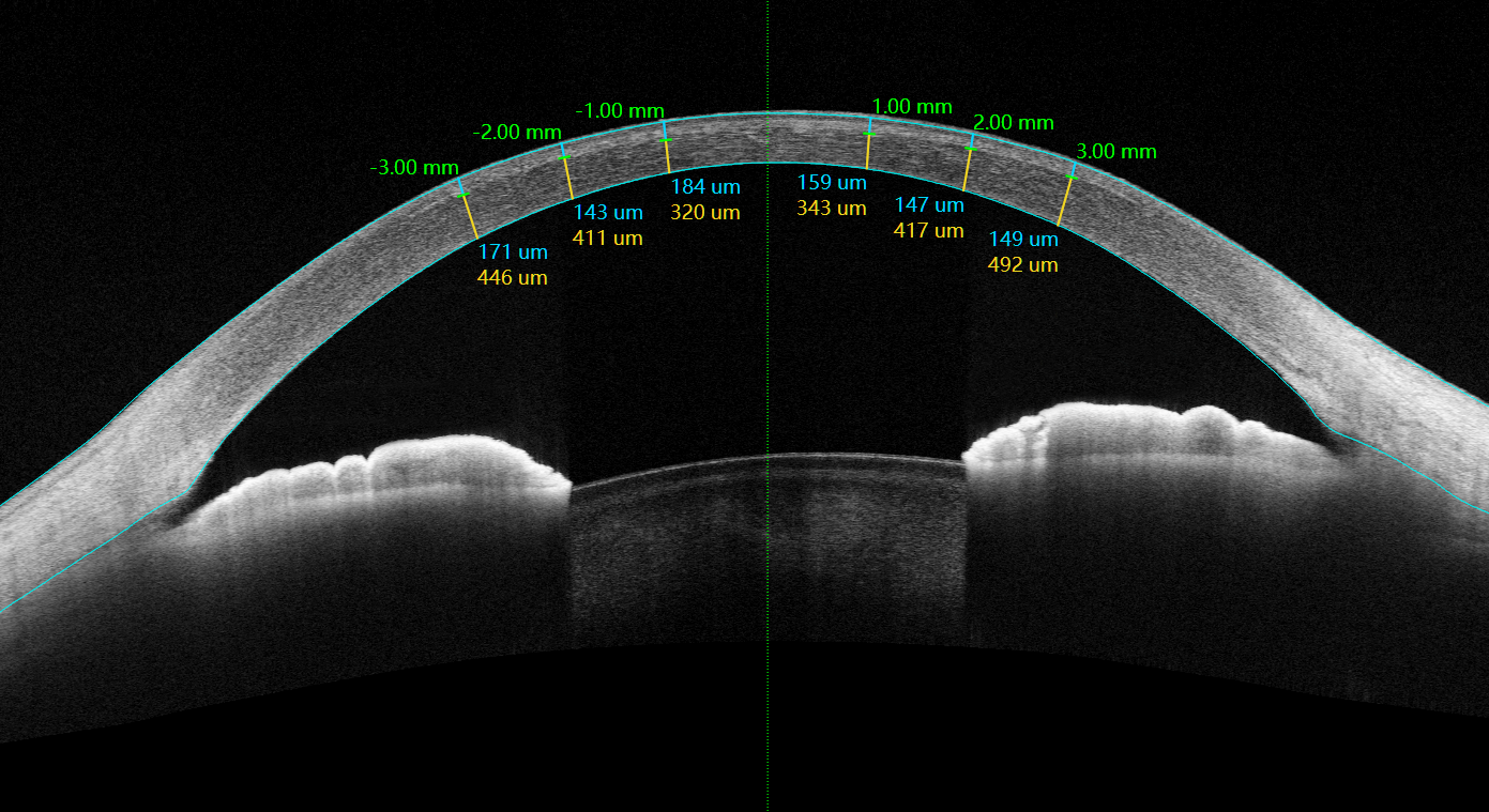
Corneal flap thickness measurements after LASIK
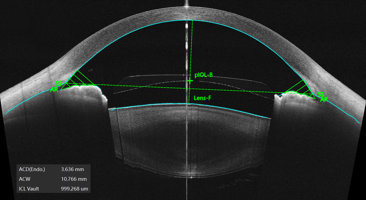
ACD and vault measurements after ICL implantation
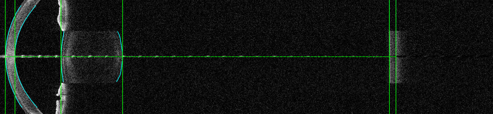
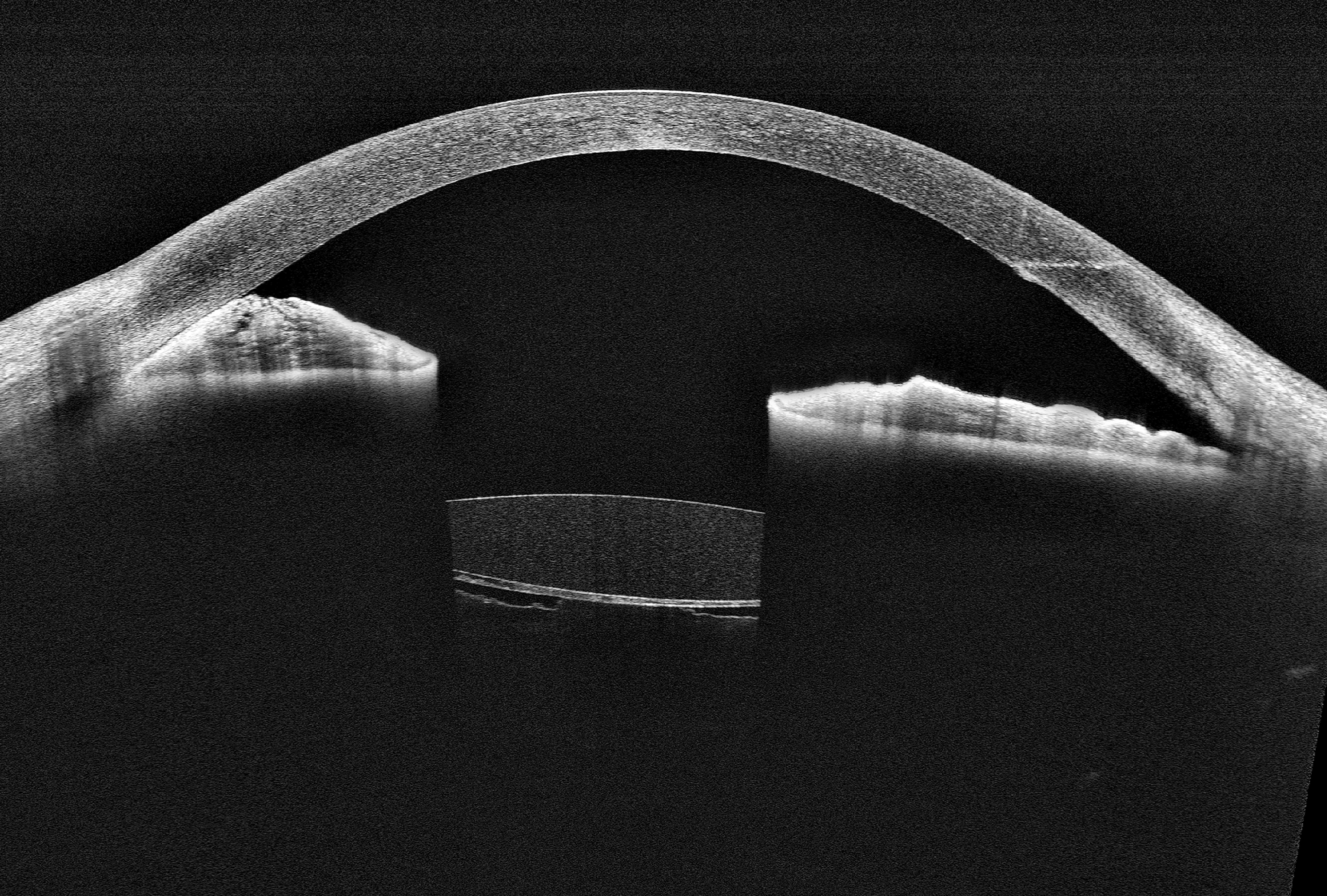
Super-Depth of AS-OCT Imaging Helps Make Surgical Plans and Assess the Results
The DREAM OCT™ could achieve a deepest imaging depth of 16.2mm (in the air) for the high-resolution AS-OCT images, which can clearly show the corneal incision, the anterior chamber angles, the lens posterior capsule, and the anterior vitreous with only one B-scan, providing a visualized monitoring tool for perioperative management, surgical planning and effect assessment.
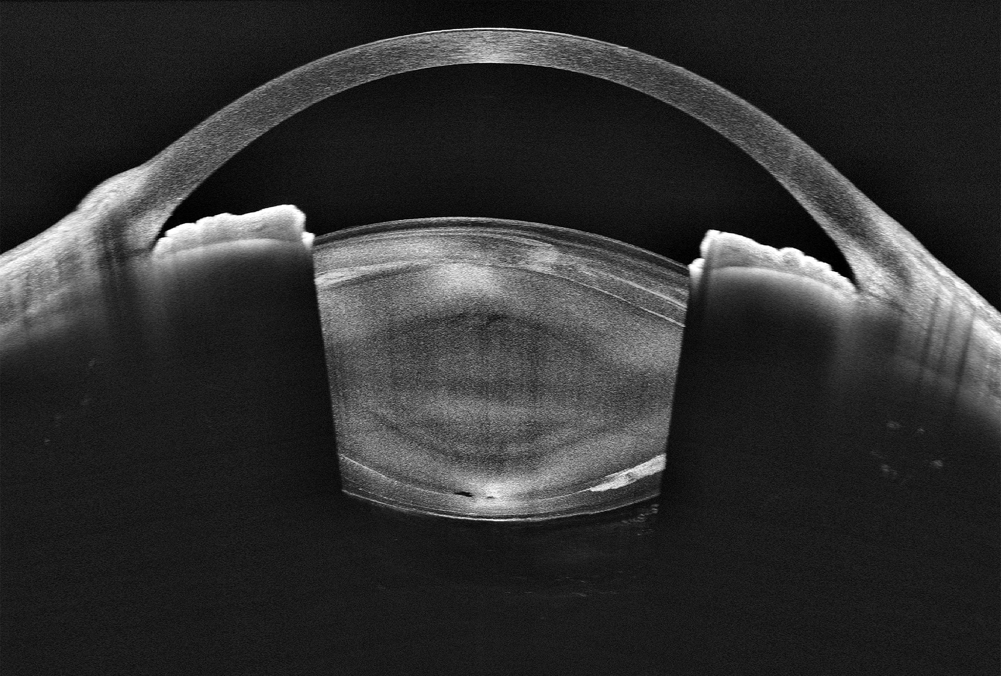
Pre-surgical examination: AS-OCT shows higher reflective signals of the lens cortex.
Post-surgical follow-up: AS-OCT shows the corneal incision, closed ACA, and close attachment between the IOL and the posterior capsule.